A new study lead by a team of researchers at the Keck School of Medicine of USC shows that younger “brain age,” a neuroimaging-based assessment of global brain health, is associated with better post-stroke outcomes. The findings could lead to better ways to predict post-stroke outcomes and offer insight on new potential treatment targets to improve recovery.
Understanding why some stroke survivors show better recovery than others despite similar damage to the brain has been a critical goal in stroke research, since it could help researchers develop better stroke rehabilitation therapies. During a stroke, blood flow to part of the brain is cut off. Without oxygen, brain cells are damaged and eventually die, resulting in brain damage known as a lesion. Studies have shown that people with similar amounts of lesion damage can experience varying amounts of recovery. Much research in the past two decades has focused on the specific location of brain damage and how the lesion affects connected networks in the brain.
This study, published April 4, 2023 in Neurology®, takes into consideration global brain health, a new way of analyzing the health of the brain based on its cellular, vascular, and structural integrity. Although global brain health has been widely examined in aging and neurodegenerative disease such as Alzheimer’s disease, it had not previously been studied in relation to stroke rehabilitation outcomes. Led by Sook-Lei Liew, PhD, of the Keck School of Medicine’s Mark and Mary Stevens Neuroimaging and Informatics Institute (Stevens INI), the team of researchers focused on a specific measure of global brain health known as brain age, which examines the biology of the nervous system through whole brain structural neuroimaging, hypothesizing that the integrity of residual brain tissue, or what is left after the stroke, may be critical for post-stroke outcomes.
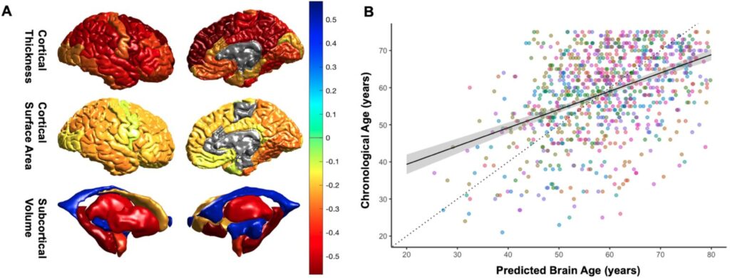
Brain age is a biomarker that predicts chronological age based on neuroimaging of structures such as regional thickness, surface area, and volumes, and is calculated using advanced machine learning algorithms, which have been widely studied at the Stevens INI. A higher brain predicted age difference, calculated as the difference between a person’s predicted brain age minus their chronological age, suggests that the brain appears to be older than the person’s chronological age. An older-appearing brain has been associated with Alzheimer’s disease, major depression, traumatic brain injury, and more.
“Brain age has not been widely explored in stroke. A lot of stroke research has focused on how damage to the brain results in negative health outcomes, but there has been less research on how the integrity of the remaining brain tissue supports recovery. We expected that younger-appearing brains would be buffered from the effects of the lesion damage and therefore have less impacts on behavior,” says Sook-Lei Liew, PhD, lead author of the study and associate professor with joint appointments at the Stevens INI, the Chan Division of Occupational Science and Occupational Therapy, the Division of Biokinesiology and Physical Therapy, and the USC Viterbi School of Engineering.
The research team conducted an observational study using a multi-site data set of 3D brain structural MRIs and clinical measures from ENIGMA Stroke Recovery, a collaborative working group of more than 100 experts worldwide who pool together post-stroke MRI data to create well-powered, diverse samples. The primary mission of the group is to create a worldwide network of stroke neuroimaging centers focused on understanding the mechanisms of stroke recovery.
The new study showed that younger brain age is associated with superior post-stroke outcomes. The researchers note that inclusion of imaging-based assessments of brain age and brain resilience may improve the prediction of post-stroke outcomes and open new possibilities for potential therapeutic targets.
“The health of your overall brain can protect you from the functional consequences of stroke. That is, the healthier your brain is, first, the less likely you are to have a stroke, and second, the less likely you are to have poor outcomes if you do have a stroke. There’s so much research on the aging brain right now, and therapeutics being developed to slow brain aging. This study ties brain aging to stroke outcomes, so any therapeutics developed to slow brain aging might also be helpful to improve outcomes after stroke,” notes Liew.
For this study, the team of experts used high-resolution MRI data from research studies. They plan to progress their brain age assessment work by applying it to routine clinical MRI data to determine if it can be an easily implemented biomarker for stroke rehabilitation outcomes. Researchers at the Stevens INI collaborate on a variety of stroke research, including the Stroke Pre-Clinical Assessment Network (SPAN), which was established to address a significant need in the scientific investigation of stroke treatment. Additionally, Liew and other USC collaborators recently released an expanded, open-source data set of brain scans from stroke patients in hopes of accelerating large-scale stroke recovery research.
For more information, and for a complete list of funding for this research, you can access the paper here.